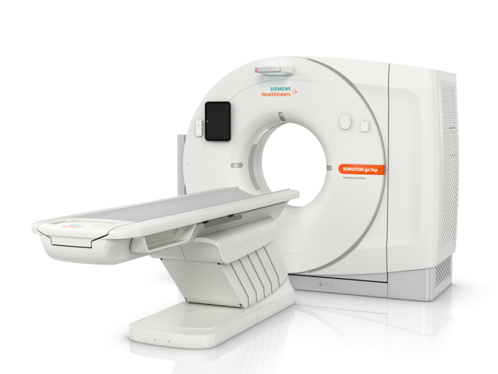
It is a world leader in image quality and scanning speed. With the Siemens SOMATOM go.Up tomograph, new technologies are introduced that are innovative for tomographs of this class:
- minimal radiation dose received by the patient during scanning;
- specialized procedures can now be performed as part of routine diagnostic practice;
allows for widespread use of screening methods.
Indications for CT
Computed tomography is performed in the following cases: for detailed planned diagnosis of a disease;
for detailed urgent diagnosis of severe trauma (craniocerebral, internal organs or large vessels); for monitoring the dynamics of the disease and ongoing therapy;
- for visualization during surgical intervention.
- Computed tomography is needed for more accurate diagnosis in the following cases: headache and fainting of unknown etiology;
- suspicion of a benign or malignant tumor of internal organs;
- pain in the spine (cervical, thoracic or lumbar region), especially with impaired mobility or numbness of certain body parts);
- severe combined trauma, TBI, spinal trauma.
Contraindications for CT
In modern diagnostic medicine, this procedure is indispensable in many cases, especially in the diagnosis of diseases associated with structural tissue changes. However, it should not be forgotten that X-rays are used in the tomograph. Despite the fact that the radiation dose from modern multislice devices is very small, abuse of computed tomography can cause harm. Even in the absence of contraindications, it is not recommended to perform this procedure more often than once every 6 months; the only exception is a vital necessity (e.g., rapidly growing tumors).
Absolute contraindications for computed tomography:
- pregnancy;
- presence of plaster casts or metal plates; mental disorders;
- severe neurological disorders; decompensated state.
Main contraindications for CT with contrast:
- hepatic and renal failure;
- thyroid disease;
- allergy to contrast agent components.
Preparation for CT
Preparation for CT of the abdominal cavity and small pelvis
On the day of the examination, wear loose and comfortable clothing, without metal rivets, zippers, and other decorative elements.
1 hour before the examination, drink 1.5 liters of still water.
When performing a diagnostic procedure with contrast – do not eat for 3 hours before the examination and drink only clean, still water.
When performing a diagnostic procedure with contrast, a creatinine test (kidney function indicator) is mandatory. If you have previously had allergic reactions to contrast agents, it is important to inform your doctor first and clarify this point when scheduling the procedure.
Preparation for CT with emphasis on the intestines (enterography)
Two days before the examination, refrain from eating foods that cause bloating.
One day before the examination, drink 2 sachets of polyethylene glycol ( “Fortrans”, “Moviprep”, “Diagnol” ) – 1 sachet at 3:00 PM with 1.5 liters of still water, 1 sachet at 7:00 PM with 1.5 liters of still water.
Perform an enema the evening before and the morning of the examination.
3 hours before the examination, have a light breakfast. Do not eat any more before the examination!
1.5 hours before the examination, take 1 sachet of polyethylene glycol ( “Fortrans”, “Moviprep”, “Diagnol” ) dissolved in 1.5 liters of still water.
In addition, you must bring the results of previously performed diagnostic procedures (discs/media), tests, medical conclusions, and a referral (if available).
What does computed tomography show?
Computed tomography helps the doctor determine the presence or absence of disease in the body. At St. Luke’s Clinic, the following diagnostic procedures are performed:
Head CT: maxillofacial region, brain, paranasal sinuses and nasal cavity, eye orbits, temporal bones;
Chest CT: chest, lungs, mediastinum;
Low-dose lung CT;
Lung CT for coronavirus infection;
Abdominal CT: liver, spleen, pancreas, stomach, colonography, enterography, adrenal glands, kidneys, ureters, bladder;
Spine CT: cervical, thoracic, lumbososacral spine,
Bone and joint CT: radiocarpal joints, hands, elbow joints, sacroiliac joint, hip joints, feet, knee joints;
CT angiography: aorta, neck vessels, upper limbs, brain vessels, lower limbs, pulmonary arteries;
Oncoscreening: search for metastases in organs, bones, lungs, lymph nodes. The specific type of study is indicated by your doctor in the referral.
Main stages of the examination:
Filling out a questionnaire with standard questions (full name, age, presence or absence of a pacemaker, pregnancy, other implants in the body, allergies, chronic diseases, etc.).
If the procedure is performed with contrast – an intravenous catheter is inserted and blood is taken for creatinine analysis.
When examining the abdominal organs, the patient drinks water before scanning in the amount prescribed by the doctor. The examination in the tomograph takes from 5 to 15 minutes. The total time spent in the clinic (with preparation and examination) takes from 20 minutes to 2 hours.
Processing of the result takes several days.
The result of the CT scan is not a final conclusion and is necessarily interpreted comprehensively for the correct determination of the presence or absence of the disease.
Duration of the study
The time you are scheduled for is not the start time of your study.
The study time depends on the anatomical area and the diagnostic tasks set. For abdominal and pelvic studies, special preparation is required before the study itself (30-120 minutes or more). A single area study without contrast in one phase can take up to 5 minutes. If several anatomical zones are studied with contrast in several phases – it lasts about 10-30 minutes. We find an individual approach for each patient, however, when additional examination is necessary, the study time may increase. Patients with urgent conditions (life-threatening) are examined first. You can stay in the clinic from 30 minutes or more depending on the complexity of the diagnostic task, which you should take into account when planning other activities or departure.
When you will receive the conclusion
After the study, the radiologist analyzes from 1000 to 5000 images, performs multiplanar and volumetric reconstructions, compares the results with previous studies (if any), which requires time. The most informative images are printed on film for the attending physician and the patient, the results of the study are fully recorded on disc. After a detailed analysis, you receive a conclusion, film, and disc with the study.
In some cases, additional time is required for the radiologist to contact your attending physician for additional clinical information about you. You can always ask the X-ray technician about the readiness time of your conclusion based on the results of the conducted study.
You receive the disc with the study recording immediately after the examination. The preparation of the doctor’s conclusion usually takes from 1 to 3 working days. For your convenience, it is possible to send the results to the specified email, messengers (Viber/Telegram/WhatsApp). The results of urgent studies are issued on the same day.